What Patients Ask Before Getting Veneers: Stains, Nightguards, and Whitening
Do veneers stain? Do you need a nightguard? Learn the facts about porcelain veneers and whitening. Schedule your consultation at Long Beach Family Dentist now!
Read More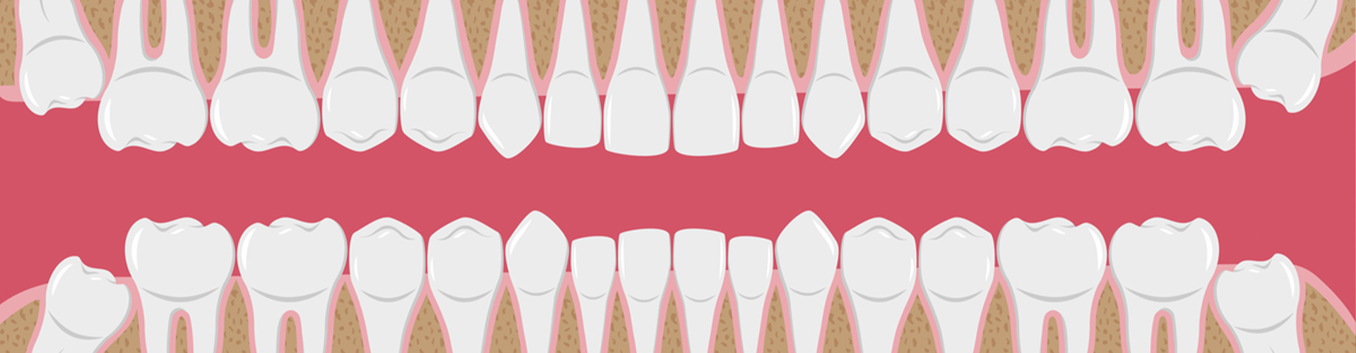
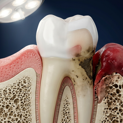
What is a tooth? A tooth can be defined as one of the structures within the mouth that allows for biting and chewing. The teeth are arranged in upper and lower arches. Teeth have different shapes, depending on their purpose. The sharp canine and the incisor tooth function in biting, and the posterior teeth or molars in the oral cavity provide grinding forces for masticating food. Areas of the tooth The parts of the tooth correspond to dental health. The tooth consists of two major parts, which are as follows: 1. Crown: The portion of the tooth which is visible in the mouth. We can divide this into an anatomical crown and a clinical crown. An anatomical crown is that portion of the tooth that is covered by enamel (hard tissue), whereas a clinical crown is the portion that is visible in the mouth. The clinical crown may or may not correspond to the anatomical crown, depending on the level of the tooth’s investing soft tissue. The clinical crown may be an ever-changing entity throughout life, while the anatomical crown is a constant entity. 2. Root: It is that portion of the tooth that is not visible in the oral cavity. This can also be divided into anatomical and clinical roots. The anatomical root is that portion of the tooth that is covered by cementum, whereas the clinical root is that portion that is not visible in the mouth. The clinical root is an ever-changing entity and may or may not correspond to the clinical root.
The tooth apparatus is more widely classified into tooth proper and the supporting tooth structure. 1. Tooth proper The tooth proper consists of enamel, dentin, and pulp. *Enamel: The hard, mineralized tissue which covers the dentin of the anatomical crown of a tooth. It is the hardest living body tissue but is brittle, especially when not supported by sound underlying dentin. It is thick on the incisal or occlusal surface and becomes feather-like at the cervical edge. It is translucent with a grayish-white color. *Dentin: The hard tissue which forms the main body of the tooth. It surrounds the pulp cavity and is covered by the enamel in the anatomical crown and by the cementum in the anatomical root. The dentin constitutes the bulk, or majority, of the total tooth tissue, but because of its internal location, it is not directly visible in the tooth. Dentin is yellowish in color and is a more resilient hard tissue than enamel. *Pulp: The living soft tissue which occupies the pulp cavity of a vital tooth. It contains the tooth’s nutrient supply in the form of blood vessels, as well as the nerve supply. Pulp consists of pulp horns (usually pointed incisal or occlusal elongations of the pulp chamber, which often correspond to the cusps or lobes of the tooth), a pulp chamber (the enlarged portion of the pulp cavity, which is found mostly in the anatomical crown of the tooth) and pulp canals (that portion of the pulp cavity which is located in the roots of the tooth) that end in an apical foramen. 2. Supporting tooth structures These structures are directly linked to proper dental health. Any injury or damage to these structures can lead to prevalent oral diseases. They are the following: *Gingiva or gums: The ‘gum’ or ‘gums’ or the fibrous tissue enclosed by the mucosal membrane that covers the alveolar processes and surrounds the necks of the tooth. The color of the gums is coral pink, which can be altered due to any oral disease such as gingivitis, causing the gums to turn to a darker shade. *Cementum: the layer of hard, bonelike tissue which covers the dentin of the anatomical root of a tooth. *Periodontal ligament: Commonly abbreviated as the PDL, it is a fibrous attachment of the tooth cementum to the alveolar bone. The PDL consists of principal fibers, loose connective tissue, blast and clast cells, oxytalan fibers, and cell rest of Malassezia. *Alveolar bone: The entire bony entity which surrounds and supports all the teeth in both upper and lower jaws or arches. The alveolar bone is the hardest tissue and is made of 45% inorganic and 55% organic matter. Purposes of the areas of the tooth The purposes of each area of a tooth are given as follows: 1. Enamel: As the outer layer of the tooth, enamel serves as a protective barrier against harmful bacteria and acids that can attack the teeth. It also protects the teeth from the pressure and stress of the teeth's daily use included in chewing, biting, and grinding. Moreover, it works as an insulator of the teeth for temperatures and chemicals that may be potentially harmful to the teeth. 2. Dentin: It provides strength to the tooth, offers protection to the pulp, provides flexibility to the tooth, and is defensive in action (initiates pulp defense mechanisms). 3. Pulp: It has the most important role in maintaining dental health. It has a formative function that helps in the formation of dentin, it is nutritive, providing nutrition by way of blood and lymph vessels, it is also sensory in action and has good defense mechanisms. 4. Periodontal ligament: Functions of PDL are supportive (provide a role in load transfer between the teeth and alveolar bone), it also has sensory action, nutritive, and remodeling. 5. Cementum: The main function of cementum is tooth support or tooth anchorage together with the principal fibers and alveolar bone. 6. Gingiva: This tissue forms a tight seal around the teeth to keep them in place and provide a barrier against bacteria. 7. Alveolar bone: It helps in protection, attachment, support, and lastly, acts as a shock absorber.
Dangers to the areas of the tooth If proper hygiene is not followed and maintained, it can compromise dental health. This can cause many diseases and other dental health issues. They are described as follows: 1. Attrition: Attrition is the loss of tooth structure or tissue caused by tooth-on-tooth contact. Although this type of tooth wear is considered part of the normal aging process, more rapid dental attrition may be due to larger dental problems. 2. Abrasion: Tooth abrasion is where the teeth start to lose enamel due to some sort of outside mechanical action; in other words, teeth are physically worn down by an external force. 3. Erosion: Erosion is the loss of tooth enamel caused by an acid attack. When the enamel is worn away, the dentin underneath is exposed, which may lead to pain and sensitivity. 4. Abfraction: Abfraction is caused by stress and pressure applied to the teeth through biting, chewing, clenching the teeth, and most commonly, teeth grinding. These forces put great stress on the teeth near the gum line, where the enamel and cementum of the teeth meet. 5. Dental caries: This is the most prevalent cause of deterioration of dental health. It is defined as the breakdown of the tooth enamel. This breakdown is the result of bacteria on teeth that break down foods and produce acid that destroys tooth enamel and results in tooth decay. In the enamel, this is caused by Streptococcus mutans and Lactobacillus acidophilus, and at the junction of cementum and enamel, it is caused by Actinomycetes israelii.
Fresh news from the dental world


Mon 8AM - 5PM
Tue 8AM - 5PM
Wed-Thu 8AM - 5PM
Fri 8AM - 5PM
Sat 9AM - 3PM
Sun Closed
Check out what our patients are saying in Long Beach, CA...


“Answers any questions...”
I been a patient here for over twelve years. Andrea greeted me and got my folder set up. Toni is very friendly and answers any questions you have promptly, when I always come in she greets you with a smile and makes your appointment enjoyable. Today Jovani and Dr. Joan made this experience great. I recommend this dentist for new patients.

“Clean and friendly...”
Very clean and friendly crew. I really enjoyed myself.

“A positive experience...”
Very clean and organized office! Entire staff was pleasant and friendly, and seems interested in ensuring I had a positive experience. Will definitely return! Affordable rates.

“My favorite dentist...”
The service here is amazing and they make you feel comfortable. I'm one of those people that hates to go to the dentist and the dentists that I went to in the past were not that great with service. Overall I found my favorite dentist service now at Long Beach Family Dentist.
“Answers any questions...”
I been a patient here for over twelve years. Andrea greeted me and got my folder set up. Toni is very friendly and answers any questions you have promptly, when I always come in she greets you with a smile and makes your appointment enjoyable. Today Jovani and Dr. Joan made this experience great. I recommend this dentist for new patients.
“Clean and friendly...”
Very clean and friendly crew. I really enjoyed myself.
Check out our accreditations, sources, and relationships in the wide world of dentistry...


Check us out and follow our Long Beach, CA practice on social media...